
Breast Health
- Shruti GOCHHWAL
- Jun 1, 2020
- 7 min read

Breast Care Health Education
It is dedicated to providing comprehensive, informative and biological education on breast health and diseases.
Breast health education starts with awareness.
Creating awareness is extremely important as in the majority of cases where a breast disease was diagnosed, patients were unaware of the symptoms or had any knowledge of the disease itself.
The impact or fatality of breast disease and disorders may vary but one thing that is clear, if left unattended, they are extremely fatal.
Moreover, being aware and looking out for early signs could prevent the disease or provide quick recovery of treated early.
Anatomy of The Breast
It is scientifically known as the mammary glands. This glandular structure consists of 12-15 lobules that produce milk when they are stimulated.
The milk is transported through a system of ducts which is joined to form bigger ducts and finally opens into the nipple. The dark skin surrounding the nipple is known as areolar.
Fats and glandular tissues are present between the lobules and duct, giving it shape and structure. It includes blood vessels, lymph vessels and lymph nodes as well.
Breast Disorders
They can be categorized into two types:
Benign Breast Disorders (non-cancerous)
Hyperplasia – It occurs when there is an increase in the number of cells in the duct or lobules of the breast and is also known as proliferation.
Although it is malignant, the risk of breast cancer is increased.
There are two types:
Atypical hyperplasia is when the overgrowth of cells develops an unusual pattern or shape.
Usual hyperplasia is when the overgrowth develops a consistent pattern.
Cysts – They are one of the most common causes of breast lump due to changes in hormone levels. Most commonly found in premenopausal women around 35-50 years of age.
They usually do not need treatment but can be drained out in case an extreme level of pain or discomfort is experienced.
They do not have any potential to cause breast cancer.
Fibroadenomas-These non-cancerous breast lumps are solid, hard and well shaped.
They are of three types:
Simple fibroadenomas are about 1-3cm in size. The cells would behave the same everywhere as seen under a microscope. In future, they would not raise the chance of breast cancer.
Some of the cells have distinct characteristics when you look at complicated fibroadenoma cells under a microscope. They raise the likelihood that breast cancer will grow marginally in the future.
Up to more than 5cm giant fibroadenoma can develop.. It characterized by its large size, rapid growth, unilaterally and manifesting as breast asymmetry or deformity of the breast.
Sclerosing adenosis- They are small growths found in the milk ducts of the breasts that occur in women from age 35-55.
It is found close to the nipples and may cause nipple discharge and discomfort. It is usually removed with surgery and does not risk cancer until it is extremely large.
Radial scars –They are usually present in conjunction with other breast disorders and are known as complex sclerosing lesions.
Radial scar is a growth of the tissues that looks like a scar when viewed under a microscope. It has a central core, which contains connective tissue fibers from which milk canals and lobules develop.
While they appear to look like mammogram breast cancers, they are not cancerous, but need no further treatment and can surgically removed if necessary.
Fat necrosis –It is in a form of the lump of the breast that is normally affected by infection, surgery or radiation treatments, or damaged or dead fatty tissue cells of the breast.
A red, bruised, and comparatively thick skin may appear around the lump.
It doesn’t require a surgery or treatment and usually goes away on its own.
Phyllodes tumors- These breast lumps are extremely fast-growing. They begin in the connective tissue of the breast and develops into tumors over time.
Phyllodes tumors are usually benign and very rarely malignant. They can be sensed physically or appear in mammograms as an irregular finding. The term ‘phyllodes’ describes how the phyllodes of tumor cells appear underneath a microscope.
Mastitis- Painful swelling of the breast when it is inflamed. This causes the breast to appear red and warm. It also occurs due to an illness and women experience flu-like symptoms.
It is common in breastfeeding mothers when a milk duct gets clogged.
The pus is usually drained out and antibiotics are administered by doctors.

Credit: unsplash caption: Cells of malignant tumors under the microscope
Malignant Disorders – Breast Cancer
The breast anatomy includes lobules, ducts and binding tissues. When either of these cells begins to grow in an uncontrollable manner it leads to breast cancer.
Breast cancer is categorized based on the specific origin of cancerous cells.
If these cells originate from the epithelial cell lining then it is known as Carcinomas.
It may also be classified by its composition, whether or not it spreads.
Invasive cancer is used to describe the types of cancers that spread and In Situ cancer is used to describe types that do not grow or spread.
Carcinoma in situ
In this type of cancer, abnormal and cancerous cells have not spread beyond the places where they were formed. This is also known as cancer of stage 0. Cancers are easily curable at these points.
Two key forms are available:
Ductal in situ carcinoma (DCIS) – It is a disease of the breast ductal tissue found in the basement membrane of the breast.
If not treated properly, 20-30% of the cases develop into invasive cancer.
Lobular carcinoma in situ (LCIS) – The secretory lobules of the breast are malignant and are found within the membrane of the basement.
Invasive Carcinoma – It is also known as invasive breast cancer. Here, the cancerous cells spread into surrounding breast tissues. It is stage 1 to 4 type of cancer.
There are two main types:
Invasive Dual Carcinoma (IDC) – is the most frequently reported form around 1 in 8 breast cancers.
The cancerous cells originate from the lining of milk ducts and spread to other parts of the body through lymph vessels and bloodstream.
Invasive lobular carcinoma (ILC) – ILCs include about 1 in 10 breast cancers. These cells originate from the milk-producing lobules and spreads rapidly from there.

Credits: pexel Caption: Breast Cancer Awareness
Breast Cancer in India
Over the past 10 years, cases of breast cancer have been steadily rising in India.
In fact, breast cancer is the number 1 cancer among women in India, over passing cervical cancer which was previously recorded.
As treatment and study on breast cancer counties to progress, the topic of breast cancer has shifted from survival through treatment to disease free survival in most western countries. This progress has also been observed in developed parts of India. However, this matter is still unknown and undertrained among the bulk of the population.
Moreover, the survival rate in India is, 60% – extremely low compared to USA which 90%. This is due to lack of awareness, screening results and late diagnosis.
In India, 25.8 women per 100000 suffers from breast cancer. Taking note that there are 1.3 billion Indians with half the female population, this will amount in up to almost 350000 women living with breast cancer and a majority of them are unaware. This is extremely alarming.
Breast Health Tips: How to Examine Your Breast
Although breast changes are not serious, it is important to examine your breasts regularly to check for growth or pain. Some changes may be caused by hormonal changes but you never know when it could be an infection or tumors.
Keep in mind early stage 0 breast cancer has the highest potential for recovery or cure as breast cancer is generally easily treated during early diagonal.
It’s essential that the moment you experience it your health care provider inspect you:
Any lumps on you breast
Continuous pain or tenderness
Fluid discharged from your nipples
Nipples get inverted
Color of the breast skin changes
To lower the risk of breast cancer, women are advised to exercise daily, keep a healthy diet, avoid alcohol and smoking, maintain good healthy body weight and target motherhood before the age of 30.

credits: unspalsh caption: how to examine your breast
Medical Diagnosis: Detecting Breast Disorders
Physical exam – The doctor will perform a physical body check mainly on your breasts and armpits to feel any lumps or abnormalities.
Mammogram- A mammogram is a special x-ray of the breast.
Breast ultrasound – A breast ultrasound is an illustration of the sound waves produced by mechanisms within your breast. It can detect both solid and liquid abnormalities.
Breast biopsy- A biopsy is the removal of a sample of breast tissues or cells that is sent to the lab for testing.
MRI-Used with magnets and radio waves for the imaging of internal breast structures.
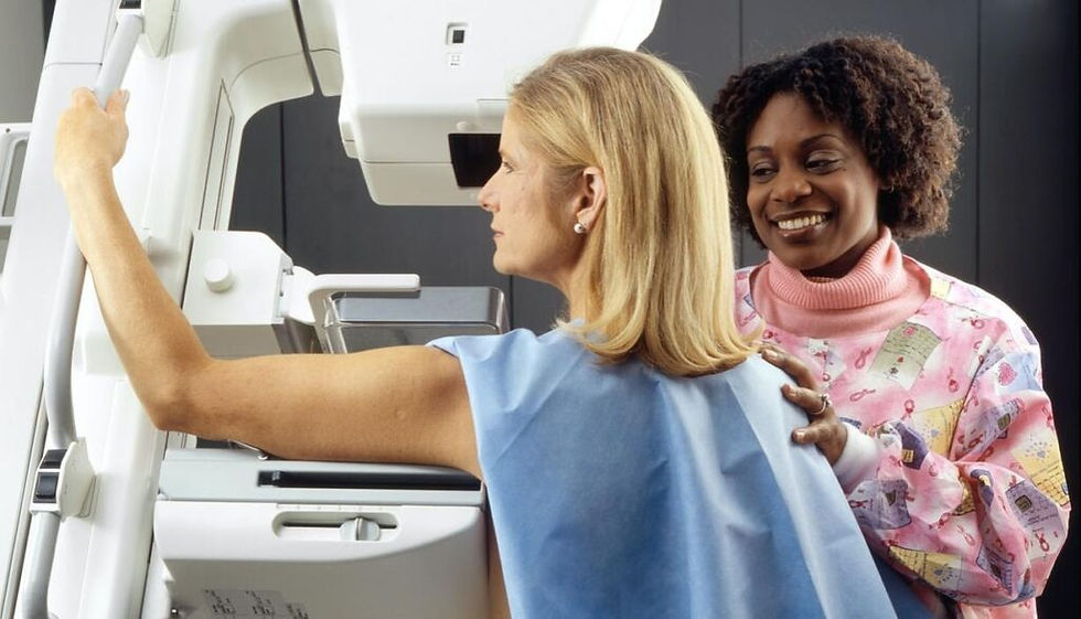
Credits : unsplash Caption: Mammogram
Treatment: Is Breast Cancer Curable?
It is not known whether or not one may be cured of breast cancer.
After medical diagnosis, doctors will determine the stage of that particular cancer. The good news is that breast cancer is very treatable in the earlier stages as surgery is easier performed when the tumor is small.
Surgery
The types of surgery include the following:
Lumpectomy – The tumor must be removed and a thin margin of healthy tissue surrounding the tumor must be cancer-free. It is also known as breast-conserving surgery as most of the breast remains but the surgery is generally followed by radiation therapy.
Mastectomy – This is the whole breast’s surgical removal. When skin can be saved, it is known as a skin-saving mastectomy, and the surgery is considered a nipple-saving mastectomy when the nipple can be retained.
Lymph node dissection- sometimes, there are cancerous lymph nodes near the breast. In this process, the surgeon removes these lymph nodes from under the arm.

Credits: Foter Caption: c
Radiation therapy
It is the use of high energy x-rays to destroy cancerous cells
It includes:
External-beam radiation therapy – The source is given from a machine outside the body.
Intra-operative radiation therapy- This is when radiation treatment is given using a probe in the operating room.
Brachytherapy- This therapy is given by placing radioactive sources into the tumor.


Comments